Medical imaging has revolutionized the way healthcare professionals diagnose and treat patients. With a range of imaging techniques available, each method offers a unique view of the body’s internal structures. A creative and fun way to explore these differences is by applying three common imaging technologies—X-ray, MRI, and CT scan—to an everyday object like a banana. This comparison not only helps visualize how each imaging method works but also highlights the distinct advantages and capabilities of each technology.
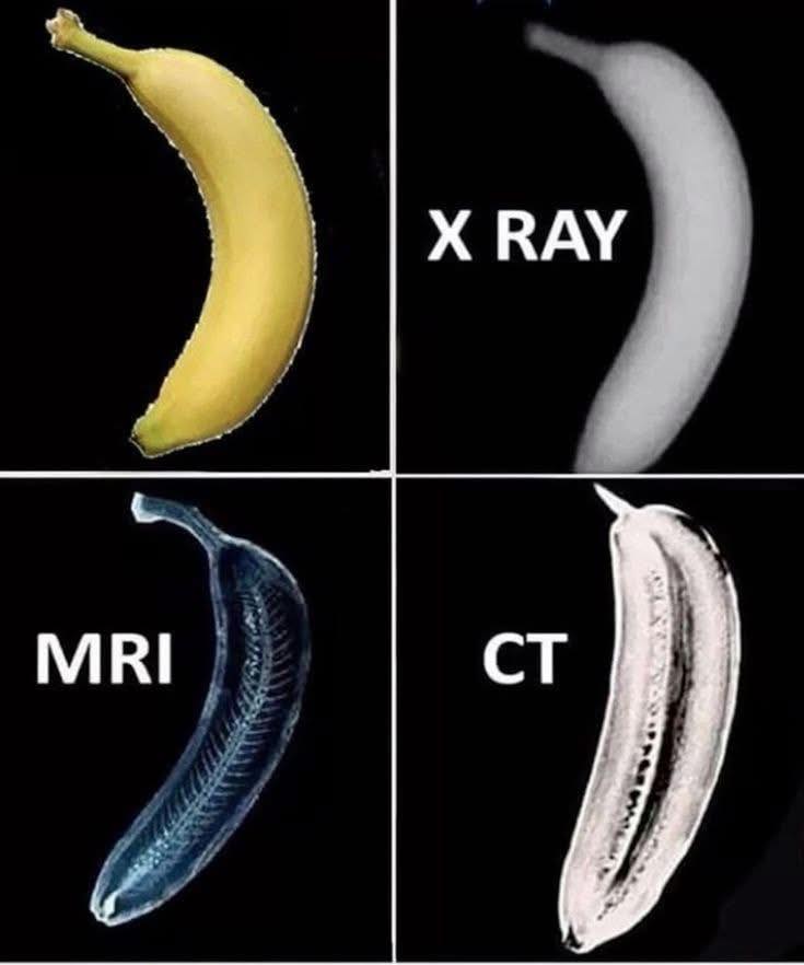
An X-ray is the most commonly known medical imaging technique, and when used on a banana, it produces a basic, density-based outline of the object. X-rays work by passing a controlled amount of radiation through the body, which is absorbed by tissues to varying degrees. Dense materials, like bones, absorb more radiation and appear white on the image, while less dense materials, such as soft tissues, absorb less and appear darker. In the case of the banana, the X-ray reveals a simple black-and-white image with only basic information about the object’s overall structure and density. The resulting image is highly useful for detecting fractures, infections, or other abnormalities in bones and tissues.
In contrast, an MRI (Magnetic Resonance Imaging) offers a more detailed and refined view of the internal structures, particularly focusing on soft tissues. When applied to the banana, the MRI produces an intricate and highly detailed image of the internal composition, revealing the subtle variations in tissue structure. MRI uses a strong magnetic field and radio waves to generate images of organs, muscles, and other soft tissues without the use of harmful radiation. The resulting image from the MRI of the banana would show not just the surface but also the intricate arrangement of cells and the texture of the banana’s internal structure, making it ideal for diagnosing conditions related to soft tissues like the brain, spinal cord, and muscles.
On the other hand, a CT scan (computed tomography) provides a cross-sectional view of the object, offering a more comprehensive perspective than X-rays. The CT scan works by combining multiple X-ray images taken from different angles and using computer processing to create detailed cross-sectional images of the body. When applied to the banana, a CT scan would create a series of slices, allowing one to see the internal structure in layers. This method provides more detail than a standard X-ray, particularly when it comes to detecting soft tissue abnormalities or even small tumors. For the banana, the CT scan would allow a closer look at the soft tissue and internal anatomy in a way that X-rays cannot.

This unique representation of medical imaging techniques through a simple banana not only demonstrates the core differences between X-ray, MRI, and CT scan but also underscores the importance of these technologies in the field of radiology and diagnostics. While each technique serves a different purpose, they are all essential tools that help medical professionals diagnose and treat various health conditions. Advances in medical imaging have greatly enhanced our ability to detect, understand, and treat illnesses, ultimately improving patient care.
In summary, using a banana to compare these three imaging technologies serves as an accessible and creative way to showcase how each method visualizes internal structures. The X-ray offers a basic outline based on density, the MRI delves into detailed soft tissue structure, and the CT scan provides cross-sectional views that emphasize the strengths of each scanning technology in healthcare.